Tumor-specific metabolic alterations have been increasingly recognized as a driving force of tumor growth.
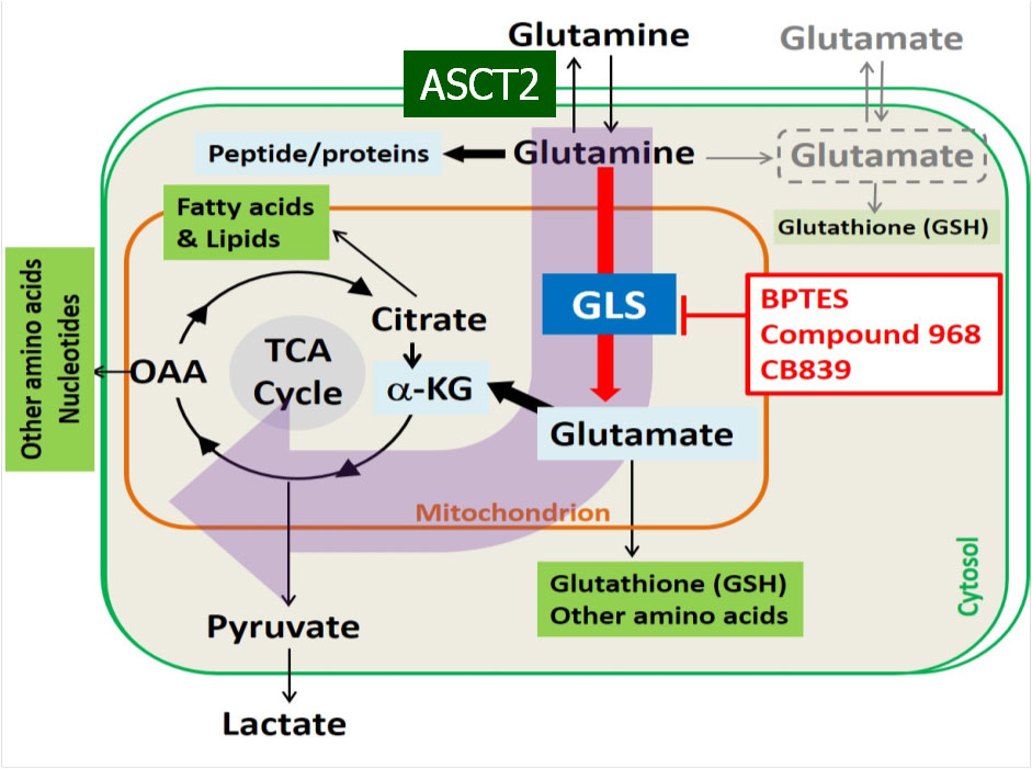
It has been discovered recently that many aggressive cancers are addicted to glutamine and deploy the glutaminolysis pathway (Figure 1) to drive energy production and biosynthesis. To exploit this unique metabolic pathway as a therapeutic target, new drugs that inhibit key steps in glutaminolysis have been developed and some have advanced to early phase clinical trials.
Biomarkers are not currently available that can quantify cancer’s glutaminolysis activity and measure its change in response to treatment but these markers are highly desirable for implementing and developing this therapeutic strategy.
Figure 1.
This project aims to develop and validate quantitative analysis tools for [11C]Gln (L-[5-11C]Glutamine) and [18F]Fluoroglutamine ((2S,4R)-4[18F]Glutamine) PET using human breast cancer xenografts.
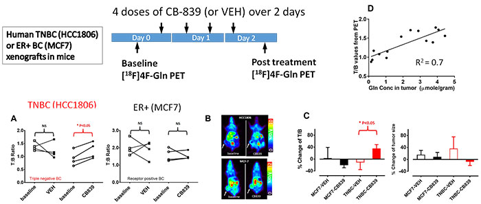
Based on our preliminary studies, we have designed studies to test the central hypothesis that “Distribution volume of [18F]Fluoroglutamine is a marker of tumor glutamine pool that is an indirect measure of glutamine metabolism (Figure 2), while [11C]Gln flux through enzyme glutaminase is an authentic marker of tumor glutaminase activity”.
Figure 2.